Humans have traveled in space for over 65 years. After the initial orbital flight in 1961, continuous crews have lived aboard the International Space Station. Future missions will require reliable medical equipment to maintain crew health during long-duration travel.
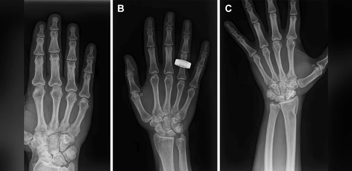
Astronauts have now produced the first diagnostic X-ray images of human bodies while in orbit. The findings appear in the journal Radiology.
For more than 40 years ultrasound served as the sole imaging method available in space. It uses sound waves and is compact, yet demands extensive operator skill. X-ray systems need a source, detector, and steady positioning, which proved difficult in weightlessness.
Recent reductions in equipment size led researchers to test compact X-ray units. A 2022 parabolic flight demonstrated brief functionality. The next test occurred during the Fram2 civilian mission, a short polar orbit lasting 3.5 days.
The crew received brief training and operated a wireless digital generator. They imaged a test object, a watch, hands, arms, chests, abdomens, and pelvises before and during flight. Radiologists on the ground rated all images as diagnostically acceptable.
Positioning subjects and hardware proved the main challenge. Hands and arms yielded the clearest results. Torso images were slightly lower in quality yet remained usable.
The same device could also examine spacecraft components for internal faults. Limits remain, including restricted imaging time and lack of real-time Earth support on distant missions. Artificial intelligence may later assist quality checks.
The unit suffered minor damage upon return but stayed operational. Future lunar or Martian systems will require greater durability.
Reliable onboard diagnosis grows essential as crews prepare for extended voyages beyond Earth.