

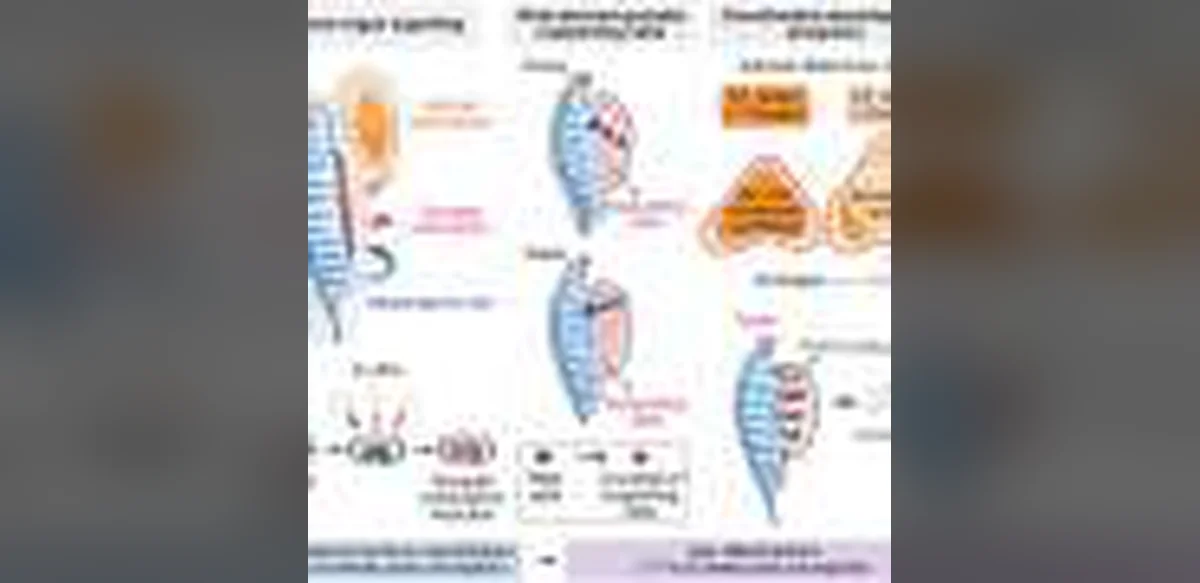
A recent investigation has produced a comprehensive spatial atlas of how human urogenital structures form during the earliest phases of embryonic growth. The work focuses on the coordinated progression of organs such as the kidneys, bladder, and reproductive tissues, highlighting the role of local cellular interactions and chemical signals that guide their simultaneous maturation.
The urogenital system arises from a shared embryonic foundation known as the intermediate mesoderm. As development proceeds, distinct regions differentiate into urinary and reproductive components. The study demonstrates that these regions do not develop in isolation. Instead, neighboring cell populations exchange molecular cues that synchronize growth patterns and tissue boundaries.
Investigators employed advanced spatial profiling methods to examine preserved human embryonic samples. This approach preserved the original three-dimensional arrangement of cells, allowing researchers to observe how gene activity varies across precise locations. The resulting dataset reveals gradients of signaling molecules that appear to direct the migration and specialization of progenitor cells.
One key observation concerns the close physical proximity between developing kidney structures and the early reproductive tract. Signals originating from one tissue appear to influence the patterning of the other, ensuring proper alignment and connectivity. Disruptions in these exchanges could underlie certain congenital conditions affecting both urinary and reproductive functions.
The analysis also identified distinct cellular neighborhoods within the developing bladder region. These neighborhoods exhibit unique combinations of active genes that correspond to different stages of organ shaping. Communication between these neighborhoods helps establish the layered architecture of the bladder wall.
Beyond descriptive mapping, the research underscores the dynamic nature of organogenesis. Rather than following a rigid sequence, development involves continuous feedback loops among adjacent tissues. Such loops allow the embryo to adjust growth in response to local conditions.
The findings carry implications for regenerative medicine and developmental biology. Understanding the spatial logic of normal organ formation may inform strategies to correct anomalies detected in prenatal screening. It could also guide efforts to engineer functional tissues in laboratory settings.
The study emphasizes the value of examining human samples directly rather than relying solely on animal models. While many developmental principles are conserved across species, human-specific timing and spatial arrangements differ in important ways. The spatial atlas provides a reference point for future comparative work.
Further research is expected to expand the atlas to later developmental stages and additional organ systems. Integrating these maps with functional studies of individual signaling pathways will help clarify how molecular instructions translate into anatomical structures.
Overall, the project illustrates how modern spatial technologies can illuminate long-standing questions about human embryogenesis. By preserving context and revealing intercellular dialogue, the work offers a clearer picture of the processes that shape essential organs before birth.